Def. Morphogenesis: generation of tissue shape that form organs and bodies
Def. Cell Differentiation: generation of the diff. cell types in tissues
It begins with division of cells so that there are enough cells to work with —> cells start to specialize because of a change in expression of genes —> cells signal to one another and also move to diff. places
Multicellular development is studies in mice, frogs, Drosophila, plants and C. elegans
Advantage for using these: rapid generation time, can be closely applied to human health —> there are many conserved evolutionary processes that are common across diff. species.
1) Development continues in adults for maintaining tissues.
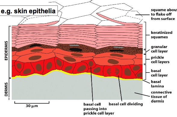
E.g. skin, there are multiple layers of epithelia that are continuously renewed. The topmost layer skin is dead and sheds constantly, then stem cells from the bottom divide and resupply the tissue.
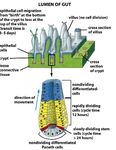
E.g. gut epithelia, the tip of the villi is constantly being replaced by stem cells on the bottom of the crypt. This process is crucial for maintaining the lining of the gut.
2) Development continues in adults for initiating new processes.
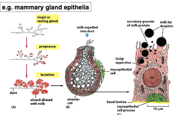
E.g. When women become pregnant, the mammary gland epithelia develops to generate milk. The duct will have many alveoli growing bigger and bigger. The epithelial layer of the alveoli make and secrete milk proteins into the duct.
The best studied for of multicellular development is embryogenesis. It has common mechanisms of morphogenesis and cell differentiation with organogenesis and stem cell development.
Embryogenesis
Haploid cells from mother and father fuse together to make a diploid zygote —> cell divides multiple times to make a loose ball of cells —> after 3 days, cells go through compaction (a process which makes cells adhere to one another) —> becomes a blastocyst
trophectoderm: outer layer of the blastocyst
blastocoel: empty space on the inside >> becomes supportive tissue that supports the embryo
inner cell mass: what becomes of an embryo
Morphogenesis requires:
1) internalization of cells
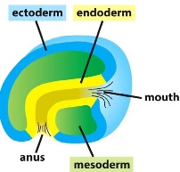
grastrulation >> determines the 3 germ layers: ectoderm, mesoderm and endoderm
e.g sea urchin
ectoderm: found on the outside >> epidermis/ nervous system
mesoderm: found in the middle >> muscle/connective tissue/blood vessels
endoderm: found on the inside >> gut/lung/liver, etc
Bending of the tissue makes digestive track, etc. Migration of cells make mesoderm.
- Mechanisms of cell internalization:
-
- ingression/delamination: individual cells separate from the early outer epithelium, and beginning to migrate (called epithelial-to-mesenchymal transition). Here the cells lose adhesion. The migratory behaviour is tightly regulated during normal development. as shown in the chick embryo experiment: cells are directed specifically to the muscle
- invagination/involution: intact epithelial sheets move inside the embryo >> stacked cells have the apical actin-filament bundles contract, narrowing the apical end, creating triangular celling. This is what makes the neural tube. (in Drosophila, the ventral expression of a transcription factor Twist regulates the invagination process)
A group of cells that are going to undergo this transition is transplanted from a quail's wing to a chick’s wing. As the wing develops, we can see that there these cells go on to make muscles.
2) elongation of the embryo
- Mechanisms of cell elongation:
-
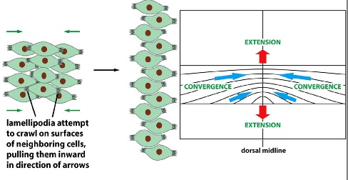
- convergent extension: cells migrate to the midline and stack on top of one another —> extension of the body
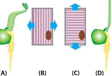
- cell division and cell shape change: e.g. root hair of the plant cells. Cell division happens at the tip so that the plant can only grow downward. e.g. orientation of cell elongation regulated by orientation of cellulose microfibrils. Here, the cells are restrained in certain directions so that they can only grow in other directions as pushed by turgor pressure. Similarly, for the gut cells, they only divide on the bottom, so they are forced to move upward.
3) fine repositioning of cells
- Mechanisms of cell repositioning cells:
-
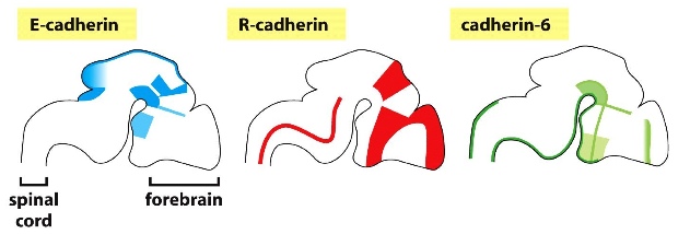
- discovered when individual frog cells were mixed together again and show that they sort themselves into different regions. Done by specific cell-cell adhesion. One type of cadherins only interact with cadherins of the same kind, so that when the cells are dissociated, cadherins look for those that they can interact with, thus creating specific zones.
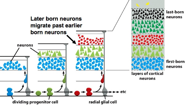
- Directed cell migration: e.g. cell migration in the cerebral cortex >> key type of cells called radial glial cells create the road where other cells can move along. Different neurons for specific regions migrate along radial glial cells and create layers above layers of neurons that can further develop into different part of the brain.
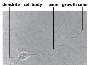
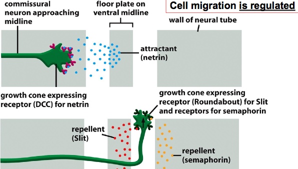
- migration of axons: growth cone moves in a path to establish the final connection. The pathfinding is very precise and can be observed by injecting dye to the neurons. e.g. connection from neural tube to brain: In order for axon to make a connection, it is attracted first to the midline when the receptors on the growth cone bind to attractant molecules —> after it goes to the midline, the growth cone expresses different receptors that bind to repellent so it moves away from the midline (two types of repp lent molecules force the axon to move in only one direction to the brain).
